The Henry H. Wheeler, Jr. Brain Imaging Center (BIC), established in 2000, is predominately dedicated to basic and clinical neuroscience using MRI methods. Dr. Mark D’Esposito, Professor of Psychology, was the initial Director of the Center and served until 2020. The current co-Directors are Prof. Jack Gallant (Neuroscience) and Prof. Chunlei Liu (Electrical Engineering and Computer Science).
Over the past 25 years over 40 different labs from many different departments and schools across campus have utilized the BIC. The BIC MRI imaging facilities have also been used to support the work of scientists and clinicians from many other institutions, both in the Bay Area and across the country.
In 2001, a Varian 4 tesla (T) MRI scanner was installed and dedicated 100% to brain imaging research, predominantly for fMRI. Berkeley’s decision to create a BIC was novel at the time in many respects. For instance, this MRI scanner was installed on a campus without a medical school and outside a clinical or hospital environment. In this way, the BIC could prioritize research without compromising clinical workload. Following our successful lead, the approach set by UC Berkeley was subsequently replicated at many other top-tier universities in the United States. The cost of the Varian scanner was entirely funded by a $5 million donation from Mr. Henry H. "Sam" Wheeler, Jr.
In 2008, a new Siemens Tim Trio 3 telsa scanner was funded by a grant from the National Science Foundation (PI: Dr. D’Esposito), and a loan from the University (50% of cost) which was paid back through user recharge over time.
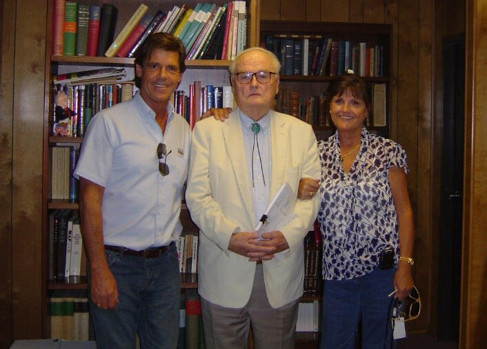
Henry H. "Sam" Wheeler, Jr. (center) with his children Hugh (left) and Nyri (right).
In 2012, this scanner was moved from a temporary facility to the Li Ka Shing Center for Biomedical and Health Sciences.
In 2020, a new Siemens Prisma scanner was purchased through an S10 grant from the National Institutes of Health (PI: Prof. Gallant), and installation was funded by UCB. This scanner replaced the older Trio scanner, which was decommissioned and sold.
The next-generation 7 tesla scanner was initially funded by two grants (starting from 2014) from NIH BRAIN Initiative (NIH R24MH106096, NIH U01EB025162) and further funded by UC Berkeley and the Weill Neurohub. The scanner is equipped with a head-only asymmetric gradient coil (200 mT m−1, 900 T m−1s−1) with an additional third layer of windings. We integrated a 128-channel receiver system with 64- and 96-channel receiver coil arrays to boost signal in the cerebral cortex while reducing g-factor noise to enable higher accelerations. A 16-channel transmit system reduced power deposition and improved image uniformity. The scanner routinely performs functional imaging studies at 0.35–0.45 mm isotropic spatial resolution to reveal cortical layer functional activity, achieves high angular resolution in diffusion imaging and reduces acquisition time for both functional and structural imaging.

